THE CARDIOVASCULAR SYSTEM: Circulation
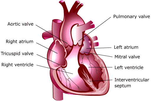
Heart Valves Blood travels through the healthy heart in one direction. Four valves control the flow through the atria and ventricles and into the pulmonary and systemic circulations. Heart valves are named for their appearance and location.
Atrioventricular valves The tricuspid valve takes its name from its three cusps, or leaflets. It is the right atrioventricular valve that connects the right atrium and the right ventricle.
The mitral valve resembles a miter- a two-cusped hat worn by bishops. It is the left atrioventricular valve that connects the left atrium to the left ventricle.
Semilunar valves
The aortic valve opens between the left ventricle and the aorta.
The pulmonary valve regulates flow between the right ventricle and the pulmonary artery.
The aortic and pulmonary valves have three cusps, and are called semilunar valves because they are half-moon shaped.
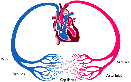
The circulatory system has two primary functions:
To deliver oxygen and essential nutrients- such as carbohydrates, fats, and proteins- to the body’s tissues
To remove cellular waste products such as carbon dioxide and urea, a nitrogen-containing end product of protein breakdown
Dark red venous blood that is low in oxygen returns to the right atrium in the two largest veins: the
superior vena cava, which collects blood from the head, neck, and arms; and the
inferior vena cava, which collects blood from the chest, abdomen, and legs. Blood within the venous system carries waste products away from the body tissues and vital organs.
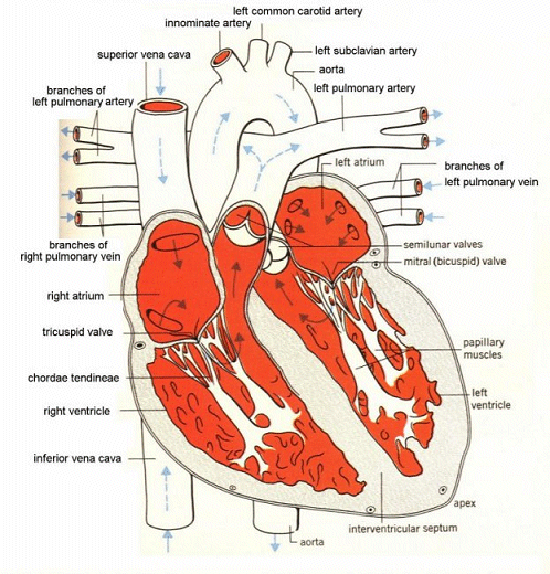
When the right atrium is filled with venous blood, it contracts and forces blood through the tricuspid valve into the right ventricle. After the right ventricle fills, the tricuspid valve snaps shut and prevents blood from flowing back into the right atrium. When the right ventricle contracts, blood is ejected through the pulmonary valve into the pulmonary arteries, where carbon dioxide is exchanged for oxygen in the lungs. Bright red oxygenated blood returns to the left atrium in the pulmonary veins.
|
Cutaway view of the human heart. |
|
|
Pulmonary vs. Systemic Pathways
Two thin-walled atria sit atop two thick-walled ventricles.
The interatrial septum separates the right and left atria.
The intervertricular septum separates the right and left ventricles.
The atrioventricular septum separates the atria from the ventricles- part of the fibrous skeleton.
The circulatory path that brings blood from the right side of the heart to the lungs then to the left atrium is the pulmonary circulation. The pulmonary circulation is the only place where veins carry oxygenated blood and an artery carries deoxygenated, carbon dioxide-laden blood. One way to remember the function of arteries is that Arteries carry blood Away from the heart; the only exception are coronary arteries, which is explained below.
When the left atrium fills with oxygenated blood from the lungs, it contracts and forces blood through the mitral valve into the left ventricle. While the left ventricle is filling, the aortic valve is closed to prevent backflow of blood from the aorta into the ventricle. Once the left ventricle is filled, the mitral valve closes and the muscle contracts, ejecting blood through the aortic valve into the aorta and systemic circulation.
The systemic circulation -the distribution of oxygenated blood to the body’s tissues and the return of deoxygenated blood to the heart- begins at the left side of the heart. From the left atrium, oxygenated blood passes through the mitral valve and enters the left ventricle. Contractions of the left ventricle pump the blood through the aortic valve into the aorta. The first arteries to branch off the aorta are the right and left coronary arteries, which bring blood to the heart muscle. Other branches of the aorta supply the head, neck, trunk, and extremities. The arteries branch into smaller vessels, the arterioles, and ultimately into a network of thousands of tiny blood vessels, the capillaries, that reaches all the body’s organs and tissues.
In the capillary bed, oxygen and nutrients leave the blood and enter cells while carbon dioxide and metabolic waste products leave the cells and enter the bloodstream. The capillaries connect arterioles to tiny venules, which empty into small veins that carry blood into larger veins and, eventually, the inferior and superior vena cavae.
|
Systole and Diastole are terms that describe the mechanical activity of the heart. Systole is the period of contraction during which the ventricles or atria force blood from chamber to chamber and through the blood vessels. Diastole is the period of relaxation during which the ventricles or atria fill with blood. When the heart contracts in the period of systole, the aortic-valve leaflets separating the left ventricle from the aorta snap upward and close off flow to the coronary arteries. Thus, the coronaries fill during diastole when the ventricles are relaxed and the aortic-valve leaflets have snapped back down. During ventricular systole, the valves between the atria and the ventricles (the tricuspid and mitral) are closed, so that blood may be ejected forward into the blood vessels. During ventricular diastole, the semilunar (the pulmonary and aortic) valves that separate the ventricles from the vessels leaving them, are closed, so that blood only enters the ventricles from the atria. The closure of these valves produces sounds that may be heard with the aid of a stethoscope. Auscultation of heart sounds permits a physician to follow the mechanical events of the cardiac cycle and to differentiate a normal pattern from one that suggests heart disease. Heart Sounds As the ventricles begin to contract, the atrioventricular valves (tricuspid and mitral) snap shut, producing a sound heard with a stethoscope as a lupp. Since ventricular systole is considered the first mechanical event in the cardiac cycle, the sound produced by closure of these valves is called the first heart sound, or S1. At the conclusion of ventricular systole, when the ventricles relax to receive blood from the atria, the semilunar valves (pulmonary and aortic) snap shut. The dupp heard with the stethoscope at this point is the second heart sound, or S2, and marks the onset of ventricular diastole. The cardiac cycle in the healthy human heart should be heard as S1/S2/pause, S1/S2/pause. The interval between S1 and S2 represents ventricular systole, and the pause represents ventricular diastole. Valvular disease produces characteristic murmurs at different points in the cycle, depending upon the site and type of the valve lesion. Other heart diseases may affect the loudness or quality of S1 or S2 or produce a third (S3) or fourth (S4) heart sound not heard in the healthy patient. |