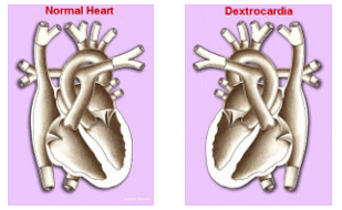
Dextrocardia and Dextroversion
Dextrocardia and Dextroversion describe a congenital condition whereby the heart is positioned in the right half of the torso. There are three conditions characterized by dextrocardia, or right-lying heart. The most common and familiar is mirror-image dextrocardia, in which the anterior-posterior relationships are normal but their right-to-left orientation is reversed. It is usually easily recognized as it is associated with some degree of abdominal situs inversus, a rare congenital condition in which the heart and major organs are transposed. The primitive loop in the embryo moves into the reverse direction of its normal position during fetal development. Approximately % 0.01 of infants are born with this disorder, and it affects males and females in equal numbers.
The second cause of dextrocardia is dextroposition, in which an otherwise normal heart is shifted to the right by some extracardiac factor. The third type is dextroversion, the least familiar and usually accompanied by other intracardiac abnormalities. In general, dextroversion consists of a rotation of the ventricular portion of the heart to the right with the atria remaining in the normal position. Usually there are transposition of the great vessels and a ventricular septal defect.
Dextroversion is a part of an exceedingly primitive arrest in development that frequently includes other thoracic and abdominal organs as well as intracardiac structures. The defect is altogether different in embryogenesis and anatomic findings from mirror-image dextrocardia. The left ventricle remains on the left, but lies in front of the right ventricle. Not only do the tips of the ventricles point to the right at the apex, but the heart is also rotated on its long axis. Since the heart is rotated to the right, the waves representing the heart beat on an ECG with conventional lead placement will indicate an abnormality. To obtain an ECG, the electrodes must also be transposed to the right side of the chest as a mirror image of the conventional setup.
DEXTROCARDIA AND SPECT IMAGING
A word of caution: although it is currently possible to acquire data in the patient with dextrocardia, there are some imaging systems that may not allow the user to process the data. Prior to scheduling a patient with dextrocardia for nuclear SPECT, be certain that your camera manufacturer’s software allows the user to manipulate the processed data for interpretation. Multipurpose single and dual head system software should offer a variety of image manipulation options (Philips-Adac Vertex, V60, Cirrus, and the like).
Obtaining an ECT of a patient with known Dextroversion requires a few changes to the imaging setup. Since the heart is in the right side of the chest, you must image from LAO to RPO. It is simple to adjust your protocol to correctly acquire and process the data in this population of patients. Lie the patient on the imaging table with his/her head at the foot end of the table, both arms up. Set up your SPECT acquisition in the computer, but edit the protocol by changing the orientation from 0 to 180 degrees. Leave the starting angle as is and scan as usual.
SPECT PROCESSING
(Philips ADAC autoSPECT + InStill 5.0) Processing a myocardial SPECT requires inversion of the reconstructed transverse slices. To do this, you must begin processing as usual, save the transverse, then go into your image manipulation under the ‘dynamic’ menu options. Then,
- Select a Frame to Invert, type in 1-19, <return>,
- Rotate Image Around the X-Axis, enter in n, <return>,
- Rotate around the Y-Axis, enter in y, <return>,
Label the inverted transverse slices then use these to reorient your slices for the non-attenuation corrected data and the attenuation corrected data. (Specific instructions should be in your protocol manual).

