
The Normal Electrocardiogram
The Normal Electrocardiogram is characterized by the wave form shown below. Each of the individual segments of the wave is designated by a letter (or
letters), and each represents a distinct phase of the cardiac cycle.
The electrodes placed on the skin are positive terminals attached to lead wires. When a wave of depolarization
advances toward a positive electrode, this produces a positive (upward) deflection on EKG. The advancing wave of positive charge in depolarization creates a positive deflection on EKG when this wave is moving toward a positive skin sensor.
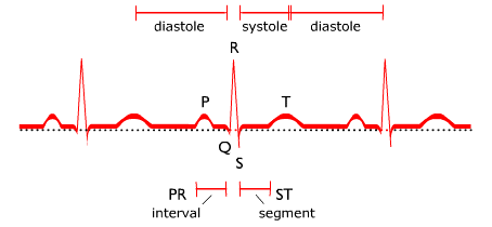
Normal ECG Shows Pattern of P, Q, R, S, and T Waves in Cardiac Cycle
P wave. The P wave represents the electrical activity associated with the impulse generation at the SA node and its subsequent spread through the atria. When the P wave is of normal size and shape, it is assumed that the impulse began in the SA node. When P waves are absent or of abnormal size or shape, it is likely that the impulse originated outside the SA node.
PR interval. The period from the start of the P wave to the beginning of the QRS complex is called the PR interval. During this interval, which normally does not exceed 0.20 second, the impulse traverses the atria and the AV node. QRS complex. The QRS complex represents depolarization of the ventricular muscle, and consists of an initial downward deflection (Q wave), a large upward deflection (R wave), and a second downward deflection (S wave). Together these waves reflect the time necessary for the impulse to spread through the bundle of His and its branches to complete ventricular excitation. The duration of the QRS complex is normally less than 0.12 second. If the duration is increased, it is an indication of bundle branch block, a condition in which the ventricles are stimulated in a delayed, abnormal manner.
ST segment. The ST segment represents the period between the completion of depolarization and the beginning of repolarization of the ventricular musculature. The ST segment may be elevated or depressed by either ischemia or infarction.
T wave. The T wave represents the normal repolarization of ventricular myocardium that occurs after ventricular contraction. Tissue injury or ischemia may cause abnormal repolarization and inversion of the T wave. The EKG records the electrical impulses that stimulate the heart to contract and gives us a valuable permanent record of electrical cardiac activity during resting and recovery periods. The electrocardiogram is inscribed on a paper strip ruled in one-mm squares. The electrocardiograph plots millivolts ( ↕ vertical axis ) over seconds ( ↔ horizontal axis ). Each vertical space represents a voltage change of 0.1 (one-tenth) mv; the voltage between two of the heavy horizontal lines is 0.5 (one-half) mv. Each small square represents 0.04 (four-hundredths) of a second: the time interval between the heavy lines is 0.2 (two-tenths) seconds in duration. By measuring along the horizontal axis, we can determine the duration of any part of the cardiac cycle. depolarization repolarization.
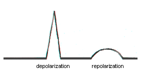
Resting heart cells “depolarize” when electrically stimulated and the negatively charged interiors of the myocardial cells become positively charged as the cells are stimulated to contract. A progressive wave of stimulation (depolarization) passes through the heart causing contraction of the myocardium. The heart remains physically quiet during repolarization, a strictly electrical phenomenon.
Electrical activity is picked up by external skin sensors and recorded as an EKG. Upward spikes are called “positive” deflections; downward spikes are called “negative” deflections. As the positive wave of depolarization within the heart cells moves toward a positive (skin) electrode, there is a positive (upward) deflection recorded on EKG. One cardiac cycle (heartbeat) is represented by the P wave, QRS complex, and the T wave. This cycle is repeated continuously. Physiologically, a cardiac cycle represents atrial systole, ventricular systole (contraction), and the resting stage between beats. Each lead records cardiac electrical activity from a different angle. The electrode pairs are different for each lead, so the tracing changes slightly in each lead as we change the angle from which we monitor cardiac activity.
The SA node begins the electrical impulse which spreads in wave fashion, stimulating both atria. The electrical stimulus originating from the SA node proceeds away from the node concentrically in all directions. This electrical impulse spreads across the atria, the atria contract, and a P wave is recorded on the EKG.
The impulse then reaches the AV node, where there is a 1/10 second pause, allowing blood to travel through the atrioventricular valves and enter the ventricles. After the pause, the AV node is stimulated, initiating an electrical impulse that starts down the AV bundle into the Bundle Branches.
The AV Bundle (Bundle of His) extends down from the AV node and divides into the Right and Left Bundle Branches within the interventricular septum. The QRS complex represents the electrical impulse as it travels from the AV node to the Purkinje fibers and into the myocardial cells causing simultaneous contraction of the ventricles.
The Q wave, when present, is the first downward stroke of the QRS complex and it is followed by the upward R wave. The upward R wave is followed by a downward S wave. This total QRS complex represents the electrical activity of ventricular contraction. The upward deflection is always called an R wave. There is a pause after the QRS complex, then a T wave appears. This pause is the ST segment, a flat piece of baseline between the QRS complex and the T wave.
Repolarization restores the negative charge to the interiors of myocardial cells. The T wave represents repolarization of the ventricles so they may be stimulated again.