
Planar Cardiac Imaging
Before Single Photon Emission Computerized Tomography (SPECT) technology became widely available, two-dimensional planar imaging was used for blood pool and all myocardial perfusion imaging. Some of the early SPECT imaging tables would hold patients up to 350 lbs., but often, obese patients found planar techniques more easy to tolerate. Nowadays, nuclear medicine imaging hardware and software have been developed to accommodate the morbidly obese patient, offering Elliptical or Circular Scan orbits. Exam tables are now built to hold 400 or 500 pounds.
In certain situations planar imaging will be your only option for obtaining a nuclear perfusion exam. Planar imaging may be acquired with gating, then summed to form composite images in three views. This is particularly useful for the morbidly obese patient, or a bedridden individual who cannot tolerate a SPECT procedure.
The morbidly obese patient will not always fit into the scan radius, even when a Circular orbit is chosen, especially when short in stature, unable to lie flat, or unable to hold arms out of the way. The newer dedicated cardiac SPECT cameras with wide, flat attenuation arms are not user-friendly for the morbidly obese patient.
PLANAR MYOCARDIAL PERFUSION STRESS/REST
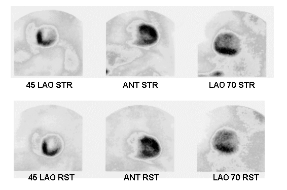
This is a set of composite images obtained with Gated Planar myocardial perfusion imaging with 99mTc-Sestamibi. The stress (top) row of images was gated, then run through a macro to composite each view into a single representative picture. The resting (bottom) row was not gated on Day 2, but care was taken to position the resting views to match the Day 1 stress scan.
The body habitus of this morbidly obese man prevented diagnostic SPECT imaging with our dual-head camera, and a gated planar exam was completed successfully with a Cirrus single-head scintillation camera. The imaging table had been replaced with an oversized gurney. Gating the images enabled our physicians to study the wall motion, although we were unable to obtain an ejection fraction.
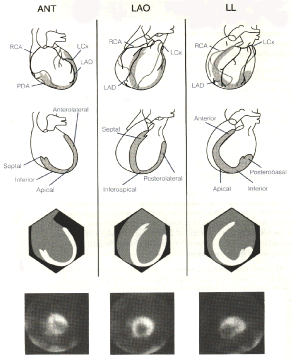
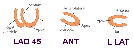
Myocardial anatomy visualized on 201-Thallium planar images
Planar imaging techniques are important to master even today. Because of the problem of overlapping structures and soft-tissue artifact inherent in this modality, it is crucial that the best counts be obtained for each view, with special care in positioning.