
Gated Tomographic Radionuclide Angiography (SPECT MUGA)
It has recently become possible to combine gated blood pool imaging with photon emission computed tomography (SPECT). It allows the automated identification of the left ventricular surface in gated tomographic radionuclide ventriculograms. Data can be displayed in three dimensions. It has been shown that the regurgitation fraction in patients with mitral and aortic insufficiency is accurately measured by gated radionuclide angiography SPECT imaging. Although the resolution of gated SPECT is not as high as magnetic resonance imaging, it is relatively inexpensive and provides an accurate picture of ventricular function.
FORMULATION PROBLEMS WITH TAGGED RBC’S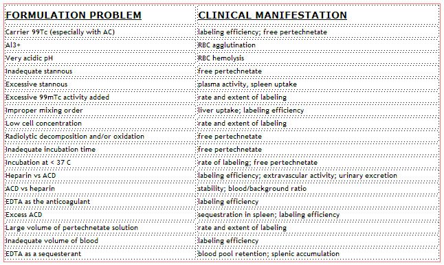
DRUG INTERFERENCE
Agents that alter, by direct pharmacological effect, cardiac function and have the potential to interfere with interpretation of the gated blood pool study:
– beta blockers (propranolol, 48 hr)
– calcium channel blockers (verapamil, 48-72 hrs)
– nitrates (nitroglycerin, 12 hr)
REASONS FOR ERROR IN EJECTION FRACTION DETERMINATION
OVERESTIMATION OF EF VALUES
- Background ROI placed too close to or over activity in spleen or descending aorta
- ROI too large (but does not include LA)
- Patients with postero-basal aneurysm
UNDERESTIMATION OF EF VALUES
- Inclusion of LA in left ventricular ROI, particularly at end diastole
- Poor separation of left and right ventricles
- Using a fixed ROI
- End-systolic ROI too large
- Inclusion of ascending aorta in end-diastolic ROI
- Placement of background ROI over photopenic area
- ECG gating problems
- Poor definition of true end-systolic volume (inadequate temporal resolution)
- Patients with antero-apical aneurysm or markedly enlarged LV
| page: |